
Groin pain when you squat or change direction? It could be Femoroacetabular Impingement Syndrome.
Have you ever felt a sharp pinch deep in your groin when you squat low, pivot on a planted leg or twist to reach something behind you? For a lot of people, this sensation becomes so familiar that it starts to feel normal.
But that pinching feeling can be a symptom of something called femoroacetabular impingement syndrome, or FAIS.
It can be a cause of hip and groin pain in active people, particularly those in their teens, twenties and thirties, and it's closely tied to the shape of the hip joint itself.
In this article, we'll walk through what's actually happening inside the hip when FAIS develops, starting with a quick anatomy refresher.
Then we’ll step through the anatomical variations of the hip that can contribute to it, who's most likely to experience it, and what can genuinely be done to manage it.
By the end, you should have a clearer picture of whether that pinch you've been feeling is worth getting looked at.
To get us started, here’s a quick anatomy session: meet your hip joint
The hip joint, known formally as the femoroacetabular joint, is what's called a ball and socket joint. Clinically, it is classified as a synovial joint.
In the hip joint, the spherical head of the femur (thigh bone) nestles into a concave socket in the pelvis called the acetabulum.
Because the ball can rotate freely within the socket, the hip is capable of three degrees of movement, which is more than most other joints in the body.
This means that the hip can accommodate flexion/extension, abduction/adduction, rotation and circumduction movements that the body makes.

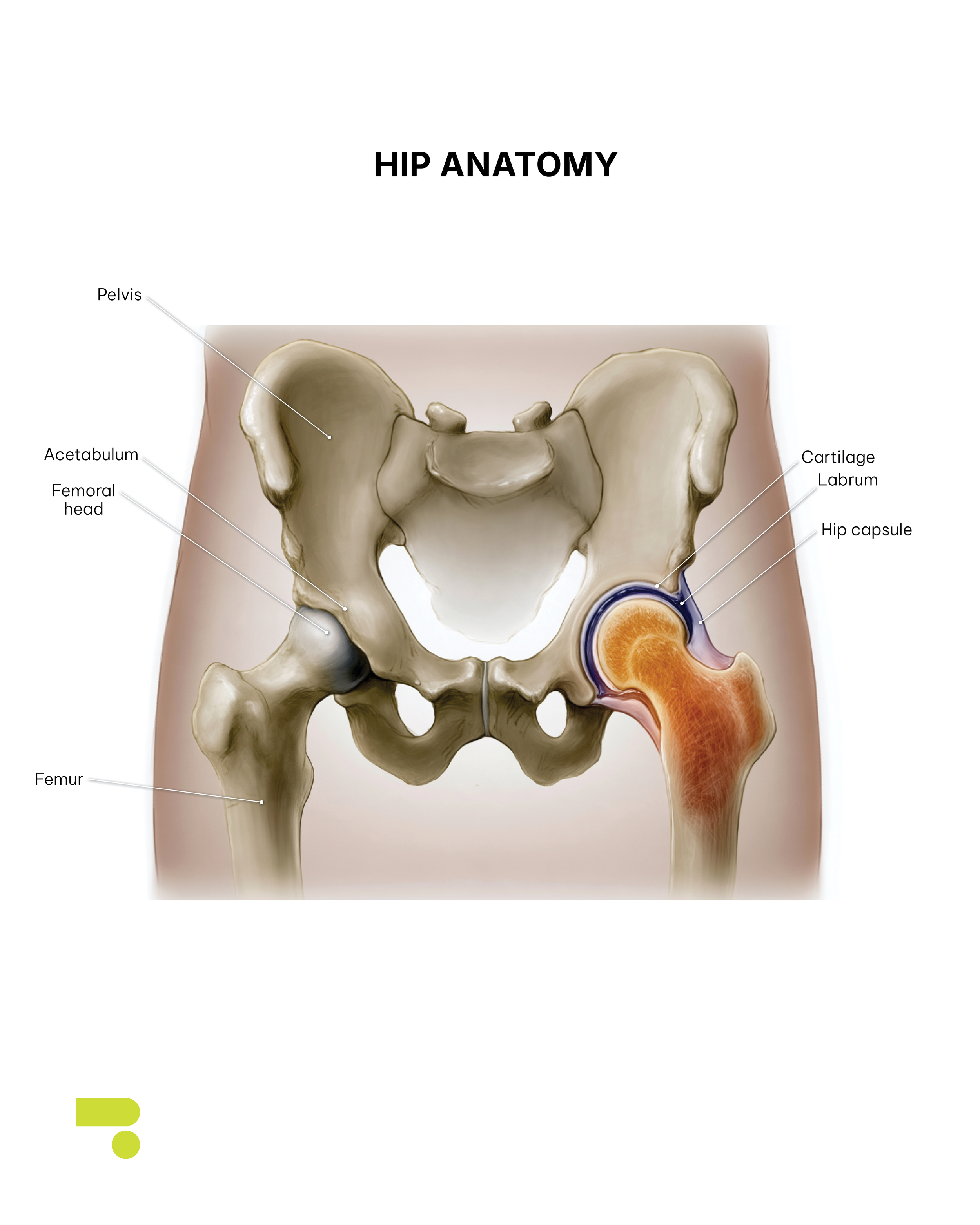
Being a synovial joint means the hip is built for exactly this kind of mobility.
It's enclosed by a capsule made up of an outer fibrous layer and an inner synovial membrane, together surrounding a fluid-filled cavity that keeps the joint lubricated.
Where the femur and acetabulum actually meet, the surfaces are coated in hyaline cartilage, which is a smooth tissue designed to let the joint glide with minimal friction while absorbing the compressive load that comes with everyday movement and weight-bearing.
This combination is what makes synovial joints ‘diarthroses’, a classification meaning they're freely mobile, more so than the body's cartilaginous or fibrous joints.
That range of motion isn't limitless, though. Two things work to keep the ball securely seated: the depth of the socket itself, and a ring of fibrocartilage called the labrum, which lines the rim of the acetabulum.
Beyond simply adding cushioning, research on hip labral function shows that it plays an active stabilising role: the labrum extends the socket's rim outward and forms something close to a suction seal where the femoral head meets the acetabulum, helping keep the joint stable through movement.
From a clinical perspective, understanding this normal anatomy matters because it's variations in the shape of this ball and socket that set the stage for femoroacetabular impingement syndrome, which we'll get into next.
So, what exactly is Femoroacetabular Impingement Syndrome (FAIS)?
The term ‘femoroacetabular impingement syndrome’ was introduced to reflect the central role of patients' symptoms in the disorder.
To reach a diagnosis, patients should have the appropriate symptoms of FAIS, positive clinical signs and imaging findings to corroborate the diagnosis.
Symptoms the patient may experience day-top-day include:
Groin pain (most common symptom)
Occasionally posterior hip pain (buttock pain)
Pain may also refer to the lateral hip, thigh, lower back, or knee
Pain is often provoked by deep hip flexion, twisting, pivoting, or changing direction
Catching, clicking, or locking sensations
A feeling that the hip or leg gives way, or buckles
Stiffness
Limited range of motion, particularly internal rotation
Clinical signs may include:
Not negative FADIR and IR@90 tests
Reduced hip muscle strength
Reduced functional task performance
Increased impingement in single leg squat
Reduced trunk function
Reduced dynamic balance
Alterations in gait
Poor range of motion
A quick note on clinical tests
Clinical tests can vary in their effectiveness, but they can help to paint a picture of what may be going on.
The FADIR test is a good example of this. FADIR stands for the different positions we can place the hip in: Flexion,ADduction and Internal Rotation.
The FADIR test is quite effective at ruling out people who don’t have FAIS.
If a person doesn’t experience any symptoms during the test, they most likely don’t have FAIS.
Though unfortunately, a positive test (i.e. reproduction of symptoms) can lead to a lot of false positives. Usually this means there are other possible explanations as to why pain is occuring in those positions.
For this reason, a test like this is said to be ‘highly sensitive’ (if you have the condition it will cause symptoms) but not very specific (it could be identifying a number of possible pathologies, we just don’t know from this test alone which one).
This is why the FADIR test alone is insufficient in diagnosing FAIS.

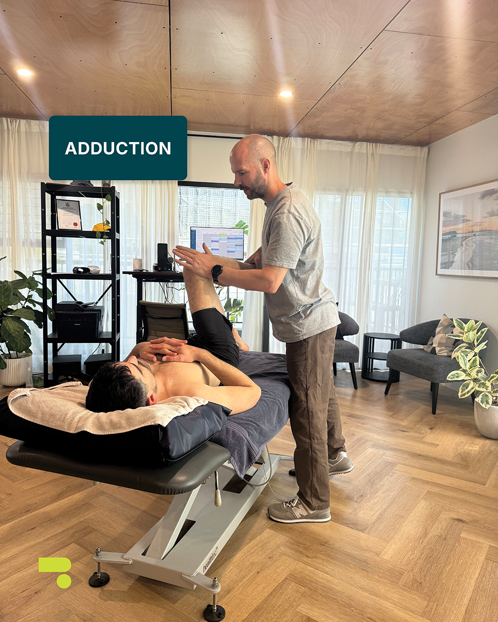

These photos demonstrate the FADIR test, which involves a combined movement of flexion, adduction and internal rotation to test for impingement symptoms.
Imaging findings should also corroborate a diagnosis of FAIS.
Plain radiographs (X-rays) should demonstrate something called cam morphology (which we will discuss further below) with increased alpha angle and reduced head-neck offset, and/or pincer morphology with acetabular overcoverage or retroversion. An MRI should be utilised when assessment of labral or cartilage injury is required.
If left unaddressed, FAIS can lead to early degenerative joint changes.
In fact, there is increasing evidence that in some people with FAIS, early degenerative changes may result in hip osteoarthritis at a later date.
To understand FAIS further, we first need to understand the different morphologies that can lead to the diagnosis of FAIS.
Meet the three hip shapes
*Note: morphology in biology is the study of the size, shape and structure of humans, animals, plants and microorganisms and of the relationships of their constituent parts. The term refers to the general aspects of biological form and arrangement of the parts of a plant, animal or human.
When considering FAIS, we need to understand the different hip shapes and how some of these hip morphologies can lead to the development of FAIS.
No two hips are built exactly the same, and the femur and acetabulum come in a handful of recognised variations.
Understanding these variations is central to understanding FAIS because it's the shape of the joint itself that determines whether impingement becomes a risk.


Normal hip morphology
In a typical hip, there's a slight concave curve (sometimes described as a ‘scoop’) where the neck of the femur meets the head, and the socket sits angled slightly forward - a feature known as anteversion.
This shape allows the femoral head to move through its full range without catching on the socket's rim.
Cam morphology
Cam morphology describes a change to this shape: extra bone forms at the front and top of the femoral head-neck junction and the femoral head loses some of its natural roundness. This often occurs around age 13 – 15.
Clinically, this is measured using something called the alpha angle, visible on X-ray or MRI. The larger the angle, the more pronounced the cam shape.
An alpha angle of greater than 60 means the presence of cam morphology. An alpha angle of greater than 78 is considered a pathological cam deformity associated with end stage osteoarthritis. An alpha angle of 83 degrees leads to 10 times higher odds of end stage osteoarthritis within 5 years.
For every degree of increase in alpha angle, the risk of end stage osteoarthritis increases by 5%.

What's striking is just how common cam morphology actually is.
Research pooling imaging data across large groups of people has found it in around two-thirds of athletes (independent of whether they had symptoms at all), in around half of people presenting with hip pain, and in roughly one in five people with no hip symptoms whatsoever.
This means that a meaningful share of the population has a cam-shaped hip and no pain to show for it.
This points to an important distinction. Cam morphology itself isn't the pathology but a structural variation that can, under the right circumstances, create excessive or repetitive contact between the femur and the socket during movement, and it's that contact that eventually produces symptoms.
Sport appears to influence how the shape develops in the first place.
Primary cam morphology, the form most often seen clinically, develops during adolescence (age 13-15) while the growth plate is still open in otherwise healthy hips. This is likely in response to, or to adapt to, repetitive high-impact loading.
Secondary cam morphology arises from prior hip disease or trauma, such as slipped capital femoral epiphysis or Perthes disease.
Pincer morphology
Where cam morphology is a femoral-side change, pincer morphology sits on the acetabular side.
It describes overcoverage of the socket over the femoral head, either in one area (focal) or around the whole rim (global) and is typically associated with either a retroverted or unusually deep acetabulum.
Mixed morphology
Many people don't fit neatly into one category. Mixed morphology describes cam and pincer features occurring together, sometimes referred to as combined ‘impingement morphologies’.
Research from a particular scientific trial found that close to 40% of people with FAIS with cam morphology also displayed pincer morphology.
These variations in the hip joint are referred to as morphologies and not deformities due to the regular occurrence of these bony variations and the absence of definitive symptoms associated with them.
These shapes turn up so frequently in people without any pain, and because their presence doesn't reliably predict who will go on to develop symptoms, ‘morphology’ has become the preferred term.
It reflects something closer to natural anatomical variation than a defect to be feared - a similar shift to how ‘degenerative change’ in imaging reports has increasingly given way to ‘age-related change’ - language that more accurately reflects that these findings may be part of the picture when someone is in pain, without being the direct cause of it.
Who gets FAIS and when?
FAIS symptoms often first appear in early adolescence, but they tend to become most pronounced in the late teens and early twenties, with peak incidence generally falling somewhere between 20 and 40 years of age.
Activities that demand an extreme range of hip motion, such as ballet, gymnastics or martial arts can bring symptoms on earlier or make them more noticeable, simply because these movements push the joint toward the end ranges where impingement is most likely to occur.
Symptoms are also more common in the athletic population generally, particularly in sports that involve rapid changes of direction, such as basketball, AFL and soccer.
Movements in these sports typically repeatedly load the hip through rotation and deep flexion.
When it comes to sex differences, overall rates of FAIS are broadly similar between men and women, though the type tends to differ: cam morphology is more frequently seen in men, while pincer morphology is more common in women.
From a clinical perspective, the timing for diagnosing FAIS matters.
Because FAIS tends to take hold during the very years someone is most active in sport, and because it can persist and contribute to hip osteoarthritis later in life, recognising the early signs in adolescence and early adulthood is where the real opportunity for management lies.
Bone shape isn’t the enemy – compression is
Having a cam, pincer or mixed hip shape doesn't hurt in itself. What actually produces pain and joint damage is when repeated compression between the femur and the socket occurs.
Repeated over months and years, particularly in someone whose sport or activity regularly pushes their hip into these end-range positions, this compression takes a toll.
Studies have found that during arthroscopic surgery on people with cam morphology, labral and chondral damage present in 90% of patients. This is a strong indication that it's the repetitive mechanical loading, not the bone shape on its own, doing the damage.
From a clinical perspective, this distinction matters. Managing FAIS isn't necessarily about fixing a bone shape that a large proportion of the population lives with symptom-free. It's about identifying and modifying the movements and loads that are driving compression in a joint that, structurally, is more vulnerable to it than most.
Why do these different hip shapes develop?
Cam morphology isn't something people are born with. It develops during childhood and adolescence, and the timing gives a clue as to why.
The extra bone at the head-neck junction typically first becomes visible on X-ray from around age 13, then continues to build until the growth plate at the top of the femur closes.
Once that growth plate fuses, the shape is set. No further change tends to occur into adulthood.
The leading explanation is that this bony growth happens as an extension of the open growth plate (femoral epiphysis) itself, occurring during a period when the bone is still actively growing and is more responsive to the loads placed on it.
This is why the theory points so strongly toward sport: young athletes going through periods of rapid growth, particularly in high-impact and change-of-direction sports, appear to develop cam morphology at meaningfully higher rates than their non-athletic peers.
Among adult athletes in high-impact sports, prevalence has been reported anywhere from around 60% up to close to 90% depending on the study and sport in question, figures well above what's typically seen in the general population.
In practical terms, this means cam morphology is largely a story of adolescence: a bony adaptation that forms in response to load during a specific developmental window, then stays essentially fixed for the rest of a person's life.
An important point here is that changes in the hip's structure and mechanics don't always announce themselves as pain straight away.
Reduced hip range of motion can occur before someone ever reports discomfort, which is why paying attention to early signs matters, particularly in young athletes still going through this developmental window.
Signs and symptoms worth watching for include:
Groin pain that comes on during activity
Noticeably reduced hip mobility
Pain or stiffness after prolonged sitting
Clicking, catching or locking sensations in the hip
Picking up on these signs early opens the door to earlier guidance around load management and monitoring, rather than waiting until symptoms are already established.
The long game: could this lead to hip osteoarthritis?
Before connecting the dots between FAIS and osteoarthritis, it helps to be clear on what hip osteoarthritis actually is.
Osteoarthritis is a progressive joint condition involving the gradual breakdown of the cartilage that normally lets the hip move smoothly and absorb load.
As that cartilage wears down, the joint loses its cushioning, movement becomes stiffer and more restricted, and pain tends to increase over time. If left unaddressed, these symptoms generally continue rather than resolve on their own.
The link back to FAIS comes down to mechanics. Every repeated instance of impingement - the compression between the femur and the socket we covered earlier - places extra stress on the same cartilage and labral tissue that hip osteoarthritis eventually erodes.
Over years, particularly in a joint that's being loaded repeatedly through the exact ranges where impingement occurs, this repetitive stress is understood to accelerate the kind of cartilage wear that defines osteoarthritis.
From a clinical perspective, this is where early management is key.
Reducing risk generally comes down to a few things: staying attentive to symptoms rather than pushing through them, modifying or reducing activities that repeatedly provoke pain, and in some cases, surgical intervention to address the underlying morphology directly.
None of these guarantee osteoarthritis won't develop, but each is aimed at reducing the cumulative mechanical stress that drives the process forward.
What can actually be done about FAIS?
Osteopathic management
The management of FAIS sits on a spectrum from conservative, hands-on care through to a range of surgical options. And where someone lands on that spectrum depends on the severity of their symptoms, the extent of any joint damage, their activity goals and how they've responded to treatment so far.
For many people, particularly earlier in the course of FAIS, conservative management is the first and often most appropriate step.
From an osteopathic perspective, this isn't about fixing the underlying bone shape but about managing the load and mechanics around a joint that's more vulnerable to compression than most.
In practice, this can look like working through a flare-up to help settle symptoms, and getting clear, honest guidance on how significant the presentation is and what the realistic trajectory might look like.
Where movement is genuinely restricted by tight or guarded tissue rather than by bone-on-bone contact, improving that mobility can help.
Building strength through the back, hip and leg supports the joint through the loads it's regularly exposed to and improving joint control, including how well someone manages deceleration and change-of-direction movements, reduces how often the hip is driven into compromising positions in the first place.
It's also worth looking beyond the hip itself. Movement through neighbouring joints affects how much work the hip ends up doing.
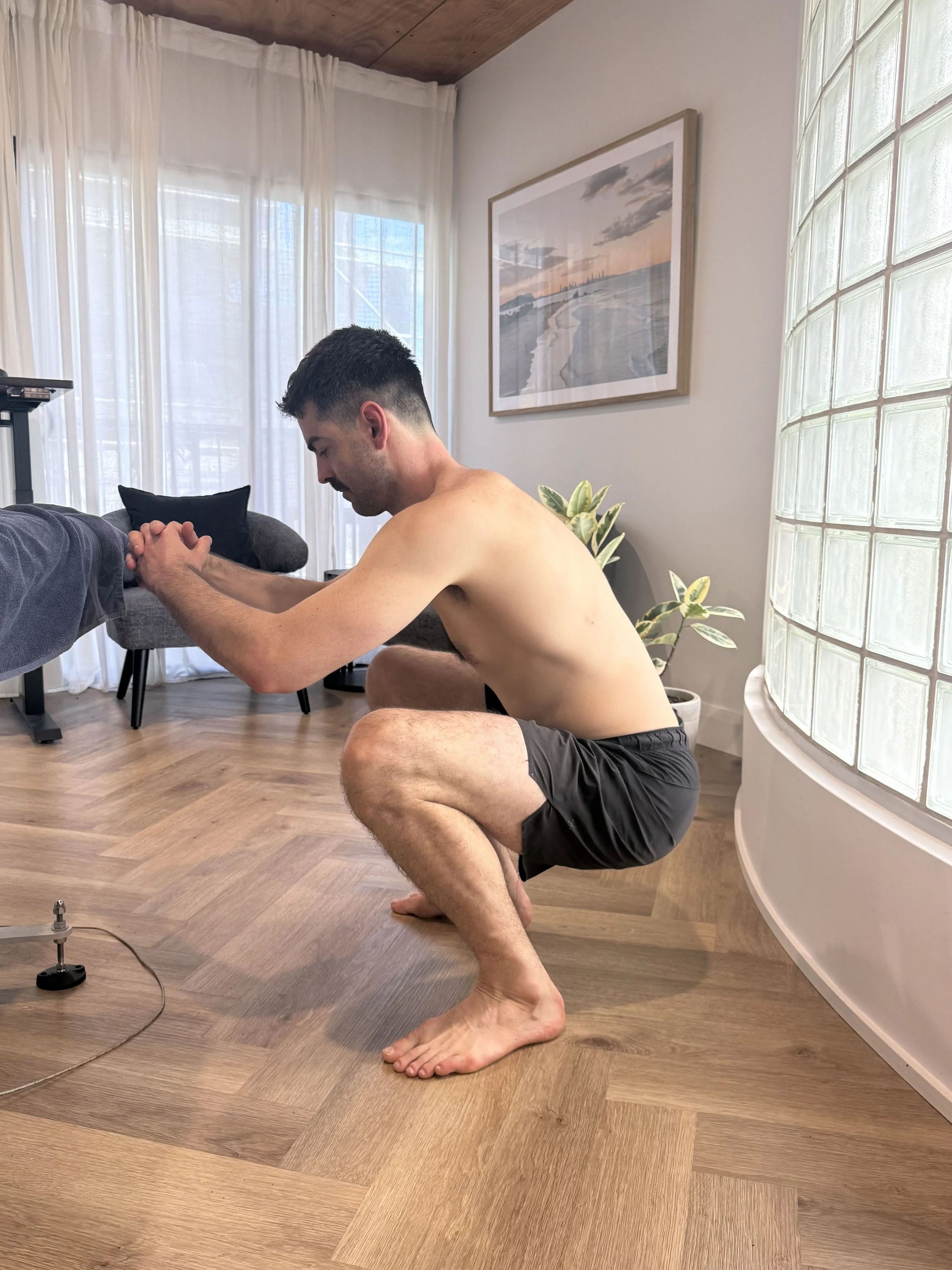
Reduced ankle dorsiflexion for example can shift additional load onto hip flexion during a squat, meaning a stiff ankle further down the chain can quietly be making a hip problem worse.
Addressing symmetry and movement quality through the back, knee and ankle is often part of a genuinely comprehensive approach.
Should early screening for athletes be considered?
Recent research suggests that early intervention for symptomatic FAIS among adolescents will lead to better long-term outcomes.
Up to 96% of adolescent FAIS patients participate in sports, underlying the potential role that athletic participation has in the development of symptomatic FAIS in this age group.
As patients age, the incidence of cam morphology increases.
It has been shown that high levels of physical activity and participation in competitive sports at a young age increases the prevalence of cam morphology in the asymptomatic population.
Participants in these sports are at a 1.9 - 8 times greater risk of developing cam morphology than non-participants.
In adolescent populations, male patients are more likely to have a cam deformity than female patients.
There is also support for a genetic component of FAIS. Siblings of cam-type FAIS patients have a 2.8 times greater risk for cam morphology compared to unrelated controls, while siblings of pincer-type FAIS patients have a 2 times greater risk of pincer morphology.
Some researchers have suggested that primary cam morphology screening could be beneficial in offering some patients preventive support at an earlier stage but screening as part of routine clinical practice may lead to overmedicalisation and overtreatment.
The general consensus for screening young athletes appears to be a no, however increased awareness around FAIS is key so that symptoms are not ignored.
When surgery becomes part of the conversation
Hip arthroscopy has gained popularity due to its potential to delay the progression of osteoarthritis. Extensive research suggests that it demonstrates long-lasting improvement and high return-to-sport rates.
Using this method, surgeons can reshape the underlying cam or pincer morphology, repair a damaged labrum and address any cartilage injury, generally with less soft tissue disruption than open surgery, smaller incisions and a faster return to sport.
There's solid evidence behind its use, though as with any surgery, complications and the need for further surgery aren’t uncommon.
Where the labrum itself is torn, repairing it rather than simply trimming away the damaged section tends to produce better outcomes, and typically involves reattaching the labral tissue to the rim of the socket with sutures.
This approach has shown strong short-to-mid-term results and generally allows a return to sport and normal activity.
More complex cases sometimes call for different approaches again. Open surgical dislocation of the hip gives a surgeon full visibility of the joint and is occasionally still used where the anatomy is too difficult to access arthroscopically, though it's more invasive with a longer recovery, and is now reserved for select cases.
Where the underlying issue includes hip dysplasia rather than impingement alone, a periacetabular osteotomy (repositioning the socket itself to improve coverage of the femoral head) may be a better fit than simply removing bone, since bone removal alone in a dysplastic hip can worsen instability.
And in people with more advanced cartilage loss or established osteoarthritis, joint preservation surgery may no longer be the right goal at all. A total hip replacement can offer more predictable pain relief and function at that stage.
There are also some adjunctive options in the mix, including intra-articular injections, which can offer both diagnostic information and short-term pain relief, and biologic treatments such as platelet-rich plasma.
Both are used in current practice, though the evidence supporting their long-term benefit is still developing.
Modern surgical management of FAIS is highly individualised but the common thread across every one of these options is the same underlying goal: restoring more favourable joint mechanics, reducing pain and protecting the joint's long-term health as much as possible.
Conclusion
FAIS comes down to a mismatch between the shape of the hip joint and the demands being placed on it.
A cam, pincer or mixed hip shape isn't inherently a problem but under the right circumstances, repeated compression between the femur and the socket can gradually damage the labrum and cartilage that keep the joint gliding smoothly.
This tends to develop during adolescence and becomes most noticeable in the late teens and twenties, particularly for people in sports involving deep flexion or rapid changes of direction.
Left unmanaged, that repetitive compression can also contribute to hip osteoarthritis further down the track.
But there's a lot that can be done and it doesn't have to start with surgery.
Conservative osteopathic management is often the right first step for many people. Where symptoms are more advanced or joint damage more significant, a range of surgical options exist too, each aimed at restoring more favourable joint mechanics and protecting the hip's long-term health.
If any of these symptoms are ringing a bell, it's worth having it properly assessed rather than working around it indefinitely.
Get in touch with the team at Form to book an assessment with Tim or Nat to figure out what’s actually going on.
References:
https://arthroscopyjournals.onlinelibrary.wiley.com/doi/10.1016/j.eats.2012.11.002
https://radiopaedia.org/articles/cam-morphology-femoroacetabular-impingement-2
https://www.sciencedirect.com/science/article/abs/pii/S0720048X15301364
https://australian.physio/inmotion/cam-morphology-fai-syndrome-and-hip-oa
https://radiopaedia.org/articles/femoroacetabular-impingement-syndrome
https://www.sciencedirect.com/science/article/pii/S0720048X26004092
https://www.orthobullets.com/knee-and-sports/3130/femoroacetabular-impingement-fai
https://journals.sagepub.com/doi/pdf/10.1177/23259671231188332
https://www.sciencedirect.com/science/article/pii/S2255497117300812
https://www.sciencedirect.com/science/article/abs/pii/S0749806312004847
https://www.sciencedirect.com/science/article/abs/pii/S1060187219300358
https://arthroscopyjournals.onlinelibrary.wiley.com/doi/10.1016/j.eats.2012.11.002